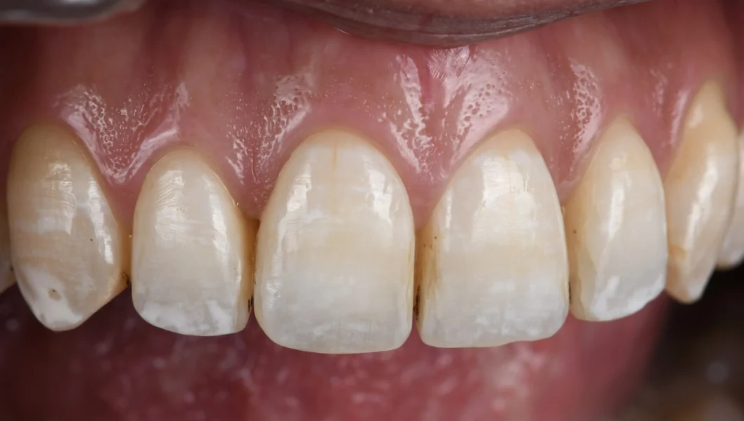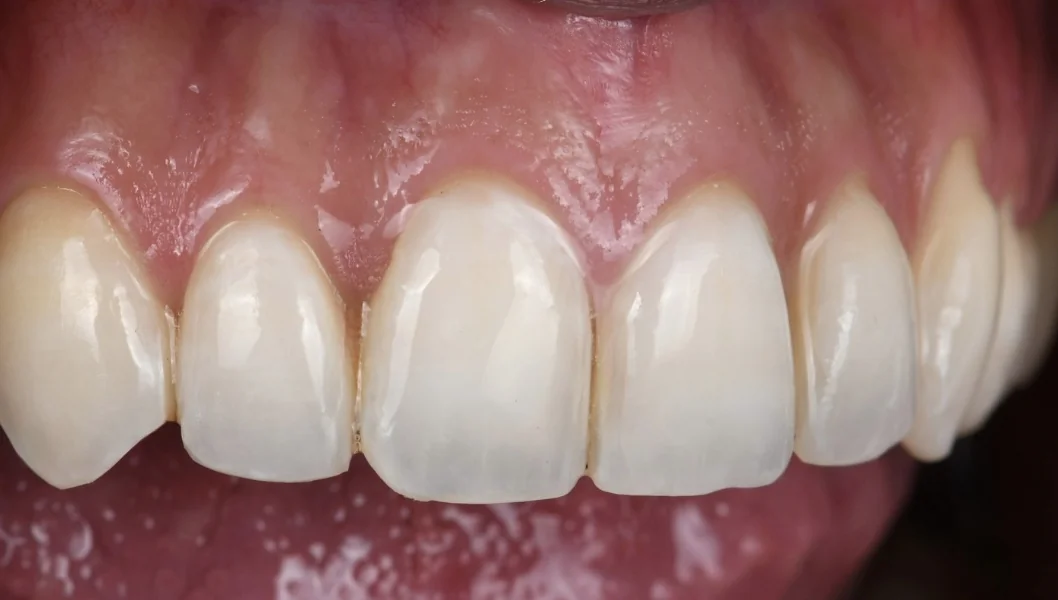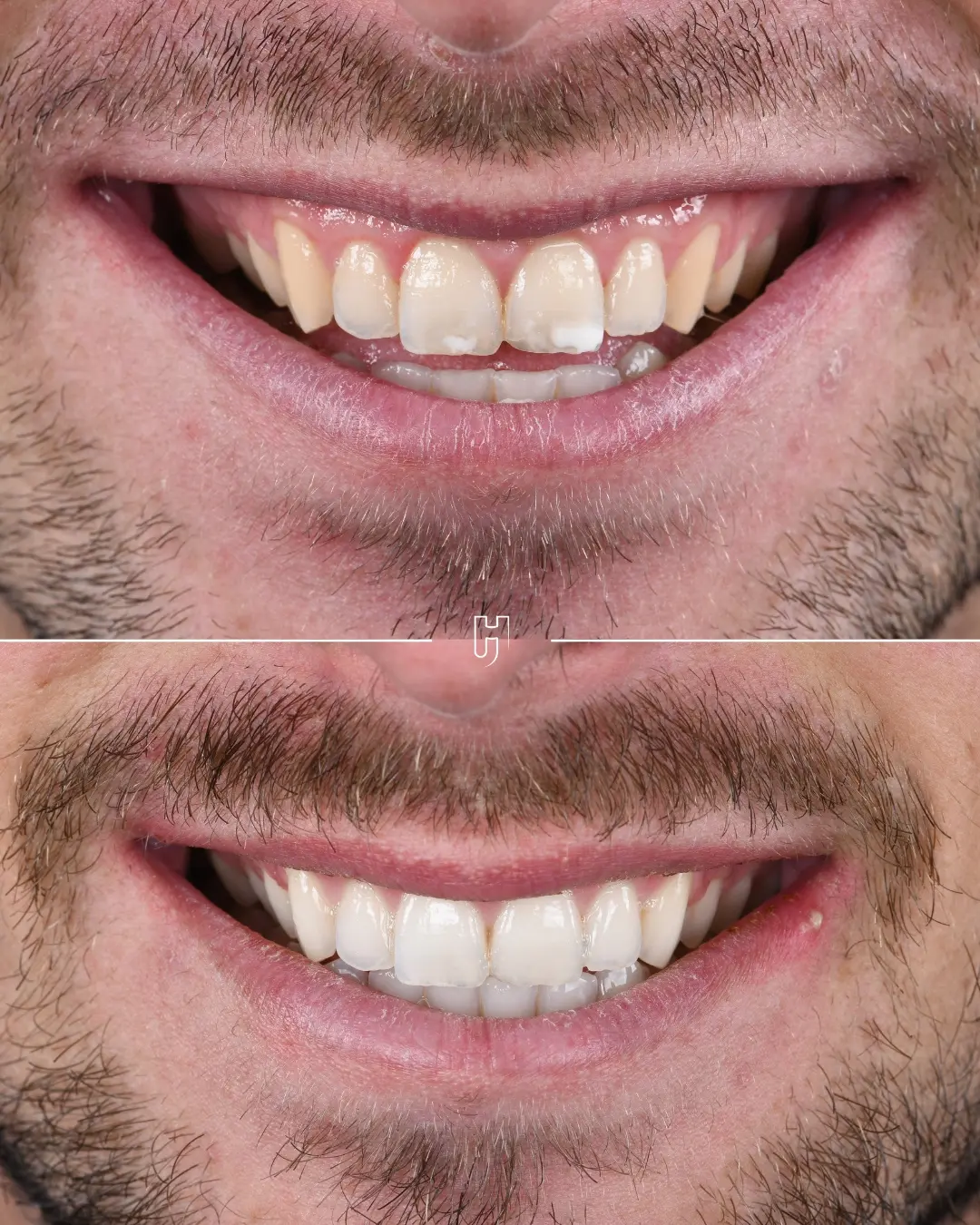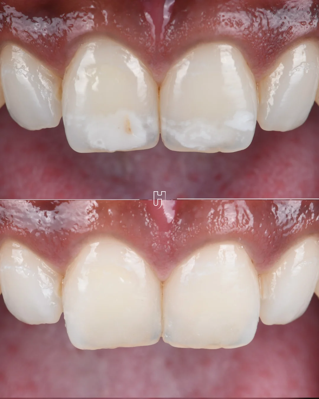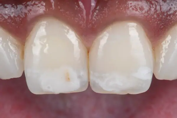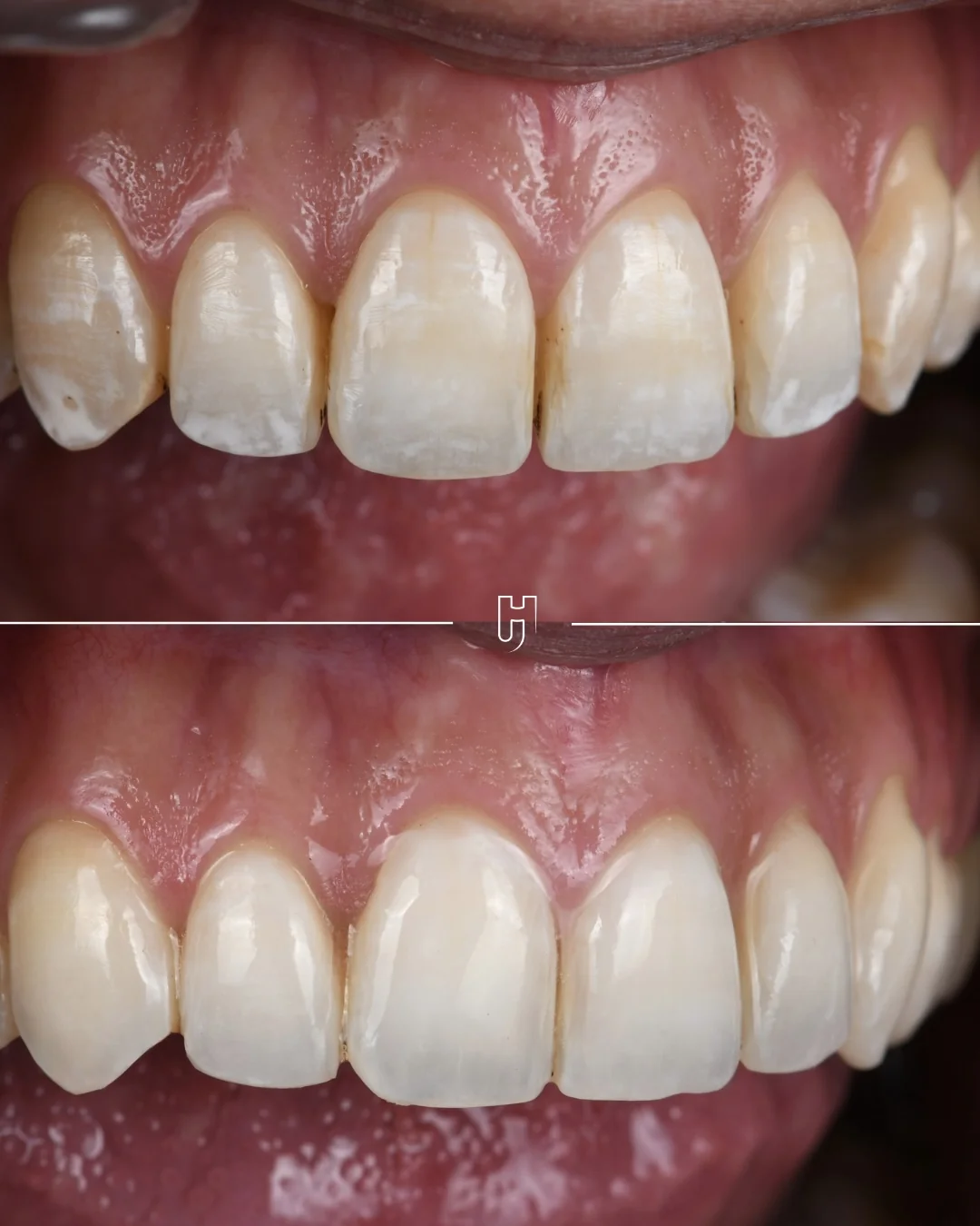
White spots caused by fluorosis
This patient had white spots caused by fluorosis, which were a significant aesthetic concern in her smile.
Dental fluorosis is linked to excess fluoride while the enamel is forming, in childhood: the spots have no sharp border and are often found on both sides of the mouth.
Reducing the contrast before masking the opacities
A white spot is all the more visible when its contrast with the shade of the tooth is strong. Hence the choice to lighten the overall shade first, to reduce the difference between the opacities and the surrounding enamel.
Infiltration comes next, only for the opacities that remain visible: each step prepares the next, the shade first, then the residual opacities.
Whitening, then infiltrating from 14 to 24
The protocol followed two steps, in this order.
At-home whitening for three weeks, to lighten the overall shade and reduce the contrast between the opacities and the surrounding enamel.
Two weeks after the end of whitening, Icon etching and resin infiltration from 14 to 24, from the upper right first premolar to the upper left first premolar, to mask the residual opacities.
Infiltration works by changing the optical properties of hypomineralised enamel. After superficial acid conditioning, a low-viscosity resin penetrates the accessible micropores by capillary action. Its refractive index, closer to that of healthy enamel than that of the air or water present in these pores, reduces the light scattering responsible for the opaque white appearance.
A uniform smile, with a conservative approach
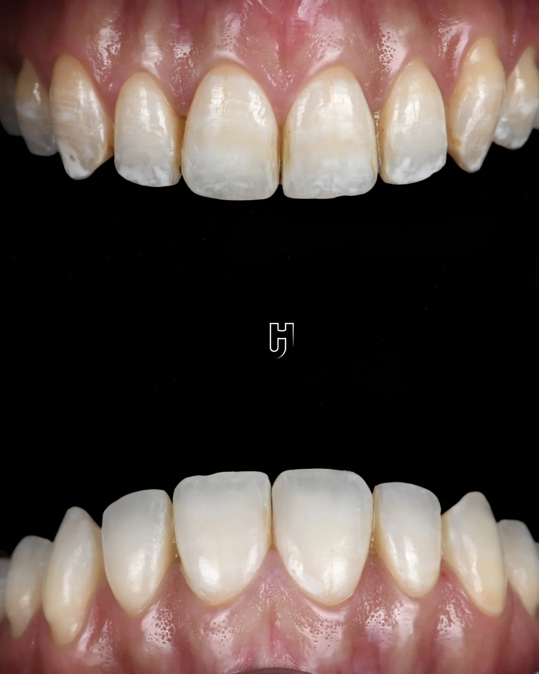
This combination made the smile more uniform while preserving dental tissue: the shade was lightened first, then the residual opacities were masked, without any restoration.
Individual result: the response depends in particular on the depth of the opacities. Another case of white spots shows a different treatment, limited to the central incisors and completed with micro-layering.